Imaging is a relatively fast, painless and cost-effective technique for viewing the internal organs and structures of the body. It can help diagnose a health problem quickly and accurately.
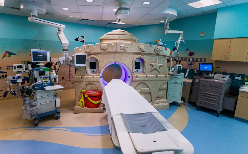
CT Scan
Computed tomography (CT) is a fast, patient-friendly form of X-ray imaging with the unique capability to view soft tissue, bone and blood vessels. CT imaging is also known as a CAT scan, or computed axial tomography.
CT imaging combines the use of a computer with a rotating X-ray device to create detailed cross-sectional images, called slices, of different organs and body parts such as the lungs, liver, kidneys, pancreas, pelvis, extremities, brain, spine and blood vessels. The procedure is fast, comfortable and painless.
Before the scan, your child may be asked to take a contrast material, either orally, through injection or both depending on the nature of the examination. The contrast material is clear and contains iodine, which makes blood vessels and structures inside the body more visible on the scanned image. Special arrangements may need to be made for children who are diabetic or allergic to iodine.
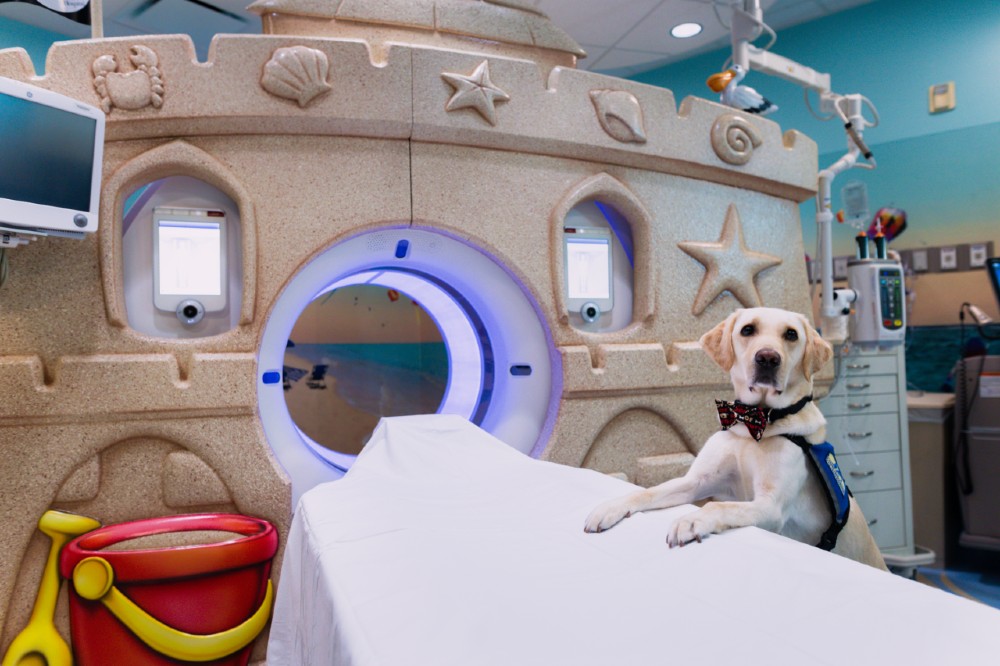
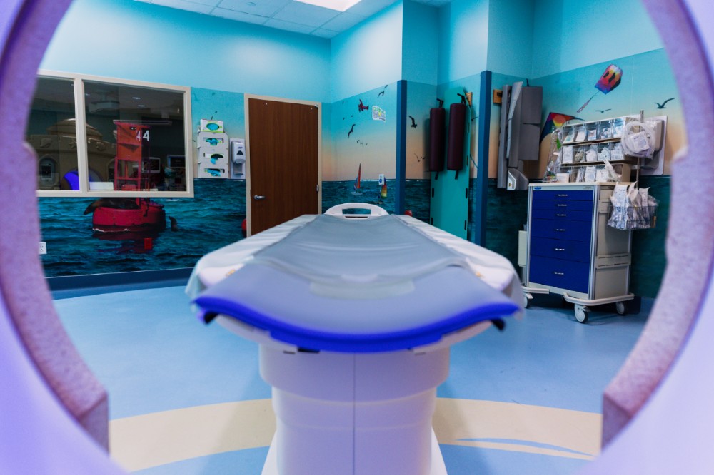
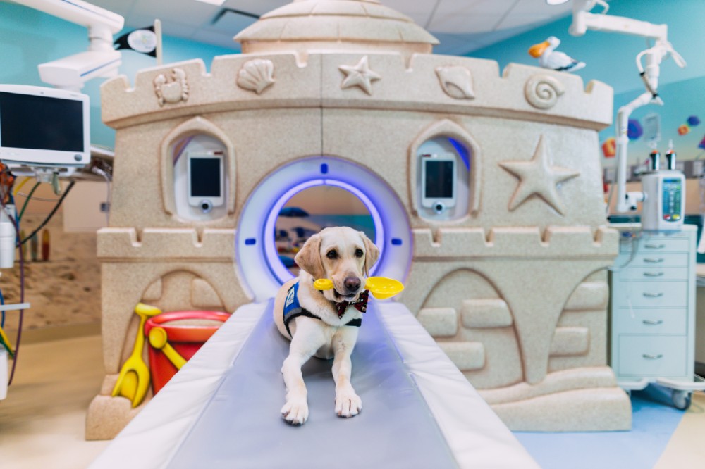
CT Scanner Image Gallery

Magnetoencephalography (MEG)
Children's Memorial Hermann brought the first MEG to Houston nearly 20 years ago.
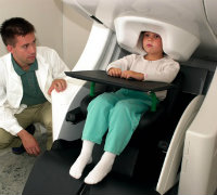
Magnetic Resonance Imaging (MRI)
Magnetic resonance imaging uses a magnetic field and radio waves to produce clear, detailed pictures of organs and other structures inside the body. MRI provides visual information about soft tissue that cannot be obtained from X-rays or other types of scans.
Because it can detect changes in normal tissue structure, MRI is most useful in providing information about inflammation, infection, tumors and injury. It can also help diagnose conditions that affect blood flow.
No special preparations are required before an MRI. However, your child will be asked to remove any metal objects before entering the scanner's magnetic field. Children with metal implants may not be candidates for MRI.
In some cases, a contrast material may be used to enhance images, evaluate blood flow and locate areas of inflammation.
Diagnostic Radiology
Our comprehensive general pediatric diagnostic services include skeletal, chest, gastrointestinal, abdominal and urinary diagnostic testing. By using the newest, safest technology, we minimize the amount of radiation needed to provide clear images of your child's internal organs and structures.
Pediatric diagnostic studies include chest, abdomen, extremity, spine and sinus X-ray; upper GI; barium enema; barium swallow; small bowel study; and IVP (kidney X-rays).
Interventional Radiology
Interventional radiology uses minimally invasive techniques to perform a variety of diagnostic and therapeutic procedures. Typically done through the skin using either a needle or small incision, the procedures are simpler, safer, more cost effective and less painful than surgery. They also require shorter recovery times.
Angiograms, myelograms, arteriograms, nephrovascular procedures and biopsies are examples of interventional radiology studies.
Ultrasound
Ultrasonography uses high-frequency sound waves and their echoes to produce images of the body. It is extensively used for evaluating the gallbladder, kidneys, liver, pancreas and blood vessels of the neck and abdomen.
The ultrasound machine uses a probe, called a transducer, to transmit high-frequency sound pulses into your child's body. To aid the transmission of sound waves, a technician will apply gel to the skin over the area of the body being examined and move the transducer over the area to visualize structures.
Nuclear Medicine
Nuclear medicine uses computers, detectors and radioactive substances to produce images of the body and treat disease. It is particularly useful for detecting tumors, aneurysms, irregular blood flow to tissues and inadequate functioning of certain organs.
Before an examination, your child will receive a radioactive tracer to make tissues visible on the scans. Bones, organs, glands and blood vessels require various types of radioactive compounds as tracers, which are either ingested by mouth or injected into a vein, depending on the type of test. The radioisotopes have very low radiation levels that decay in minutes or hours and do not harm the body.
Common uses of nuclear medicine include diagnosis and treatment of hyperthyroidism (Grave's disease), cardiac stress tests to analyze heart function, bone scans for orthopedic injuries, lung scans for blood clots and liver and gall bladder procedures to diagnose abnormal function or blockages.
To contact Children's Memorial Hermann Hospital, please fill out the form below.