Breast Ultrasound in Houston, TX
Breast cancer is one of the defining medical issues of our time. According to the National Institutes of Health, 1 in 8 women in the U.S. will develop invasive breast cancer over the course of her lifetime. The good news? Unlike some cancers, breast cancer is treatable when caught early, and the technology for early detection has improved significantly over the past few decades.
The American Cancer Society recommends women receive annual breast cancer exams starting at the age of 40. For women with certain risk factors; however, a standard mammogram may not be sufficient, and additional testing may be advised by their physician.
What is a Breast Ultrasound?
A breast ultrasound uses sound waves to produce real-time images (sonograms) of the tissues inside the breast. Breast ultrasounds can show all areas of the breast, including the area closest to the chest wall, which can be difficult to evaluate with a traditional mammogram.
Pregnant women often receive ultrasounds in order to monitor fetus development. Because sonography is non-invasive and does not use ionizing radiation, it is safer than traditional imaging procedures like radiography (X-ray) and computed tomography (CT).
Ultrasounds are used to visualize many other parts of the body as well, from muscles and tendons to internal organs. Over the past 50 years, this technology has become widely used as a diagnostic tool by radiologists and sonographers to capture the size and structure of organs, as well as the composition of any masses detected within the body. Ultrasounds are widely available, easy to operate and relatively inexpensive when compared to other imaging procedures.
Why Do I Need a Breast Ultrasound Exam?
Breast ultrasounds are typically scheduled as a follow-up to a screening mammogram. Much like Breast Magnetic Resonance Imaging (MRI), this imaging procedure is used to provide additional information about any lumps or masses detected during a self-examination or routine screening mammogram.
Breast Ultrasounds for Dense Breasts
Your doctor may also request a breast ultrasound if you have dense breast tissue. Dense breasts are characterized by an overabundance of fibrous or glandular tissue and minimal amounts of fat.
In the state of Texas, breast cancer screening providers are legally required under Henda's Law to notify women with dense breast tissue that traditional mammograms will not be as effective for them at detecting breast cancer.
Studies have shown that women with dense breasts are six times more likely to develop breast cancer. According to BreastCancer.org, 43% of women between the ages of 40 and 74 are classified as having dense breasts. If you have dense breasts, ask your physician about the benefits of breast cancer imaging procedures like breast tomosynthesis (3-D mammogram) and breast ultrasounds.
What You Should Know Before Your Breast Utrasound Exam
Before scheduling a breast ultrasound, check with your insurance provider to see if the exam is covered under your current plan. Breast ultrasounds are not currently part of the official National Comprehensive Cancer Network (NCCN) or American Cancer Society (ACS) breast cancer screening recommendations, and many insurance plans do not cover breast ultrasounds as part of a standard breast cancer screening.
While screening mammography is considered the “gold standard” for breast cancer exams, it is not as effective in detecting cancer in women with dense breast tissue. However, breast cancer detection in women with dense breasts improves by over 55% when mammography and ultrasound are used together.
At Memorial Hermann, we do our best to keep out-of-pocket costs for breast ultrasounds as low as possible.
Please bring a CD of your prior studies if your prior breast health studies were performed outside of Memorial Hermann. If you need us to request your prior films from another facility, complete a medical release of information form and return it to our facility prior to your appointment. Once received, we will request the films. Comparison with prior imaging is key to assessing for change and accurate diagnosis.
How Does a Breast Ultrasound Exam Work?
If you are scheduled for a breast ultrasound, you will prepare for an ultrasound in the same way you would a standard breast cancer exam.
To prepare for your breast ultrasound:
- Consider scheduling your breast exam one to two weeks after your period begins. Breasts are the least tender just after menstruation, making the exam more comfortable.
- Wear loose-fitting, easy-to-remove clothing. You will be asked to remove clothing from the waist up for the exam.
- Before the exam, inform your physician if you are pregnant, breastfeeding, have breast implants or are currently suffering from any medical conditions.
Your breast ultrasound will be conducted in a private exam room by a registered ultrasound technologist. Once in the room, you’ll be asked to lie down on an examination table, and the technologist will use a handheld transducer to perform the ultrasound.
Warm ultrasound gel will be applied to your breast, which helps the transducer pick up sound waves. The technologist will move the transducer back and forth over your breast until they capture images of the entire area. They may ask you to raise your arm above your head in order to obtain certain images. There will be light pressure from the transducer as it passes over your breast, but you should feel little to no discomfort.
Once the exam is finished, the technologist will wipe off the ultrasound gel. Any remaining gel will dry quickly and will not stain or discolor clothing.
How Long Does a Breast Ultrasound Exam Take?
At Memorial Hermann, we understand your time is important, and our experienced staff is committed to making sure your exam is quick and thorough. In fact, your entire breast ultrasound procedure should take no more than 30 minutes, from check in to exam completion.
When Should I Expect Results from My Breast Ultrasound?
Sonogram images from your breast ultrasound will be sent for analysis to one of our affiliated radiologists. Once they have interpreted the images, a report will be sent to your primary care physician.
Memorial Hermann’s advanced diagnostic resources and technology, coupled with the expertise of our imaging radiologists, provides a level of patient care that you won’t find anywhere else in the country – or the world.
Trust Memorial Hermann for Compassionate, Professional Breast Care in Houston
Only 65% of women say they’ve had a breast cancer exam in the past two years. If you’re a woman over 40, a mammogram and, if needed, an ultrasound, should be part of your annual breast cancer screening plan. This will greatly increase your chance of detecting breast cancer early, when the success rate for treatment is close to 100%.
When it comes to the health of you and your loved ones, you want the best care available. At Memorial Hermann, we strive to provide the highest level of patient and diagnostic care. And by offering imaging procedures at many of our locations across the Greater Houston area, we try to make it quick and easy to fit breast care into your schedule.
Clinics Offering Screening Mammograms
Mammograms are considered one of the most effective screening tools used in the early detection of breast cancer, and when caught early, breast cancer may be easier to treat. Take your health into your own hands by making a screening mammogram part of your annual breast care routine. Select a location below to schedule your mammogram online or by phone.
-
Memorial Hermann Breast Care Center at Cypress HospitalGet Directions27700 Northwest Fwy Ste 140, Houston, TX 77433
-
Memorial Hermann Imaging & Breast Care Center at Greater Heights HospitalGet Directions1635 N Loop West, Houston, TX 77008
-
Memorial Hermann Breast Care Center at Katy HospitalGet Directions23920 Katy Fwy Ste 120, Houston, TX 77494
-
Memorial Hermann Imaging Center at Convenient Care Center in KingwoodGet Directions4533 Kingwood Dr Ste 200, Kingwood, TX 77345
-
Bobetta Lindig Breast Care Center at Memorial Hermann Memorial City Medical CenterGet Directions925 Gessner Rd Ste 300, Houston, TX 77024
-
Memorial Hermann Imaging Center at Northeast HospitalGet Directions18955 N Memorial Dr Ste 100, Humble, TX 77338
-
Memorial Hermann Imaging Center - PearlandGet Directions10905 Memorial Hermann Dr Ste 104, Pearland, TX 77584
-
Memorial Hermann Breast Care Center at Southeast HospitalGet Directions11800 Astoria Blvd, Houston, TX 77089
-
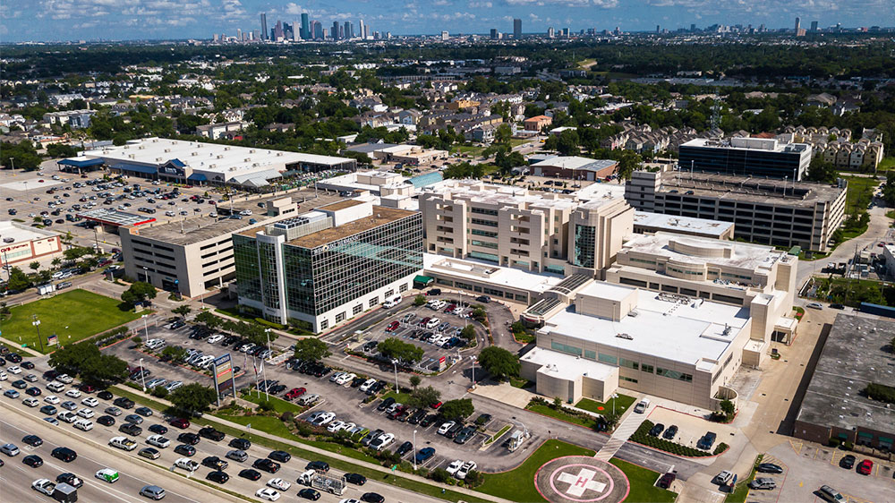
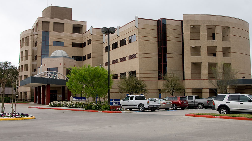
Memorial Hermann Breast Care Center at Southwest HospitalGet Directions7600 Beechnut St, Houston, TX 77074
-
Memorial Hermann Imaging Center at Convenient Care Center in SpringGet Directions7474 N. Grand Pkwy W, Spring, TX 77379
-
Memorial Hermann Breast Care Center at Sugar Land HospitalGet Directions17510 W Grand Pkwy Ste 120, Sugar Land, TX 77479
-
Memorial Hermann Breast Care Center at The Woodlands Medical CenterGet Directions9200 Pinecroft Dr Ste 150, The Woodlands, TX 77380


Contact Us
Complete the form below to be connected to our Nurse Navigator – a dedicated registered nurse who specializes in breast health and is available to provide education and resources.